Figure 4.

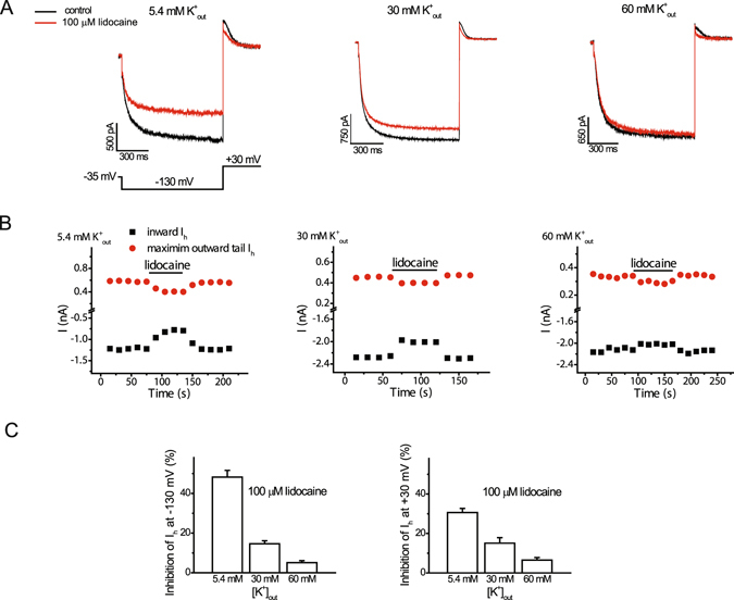
Elevation of extracellular potassium concentration opposes the effects of lidocaine on HCN1-mediated Ih amplitude. (A) Representative traces of inward steady-state and outward Ih under control conditions and following application of 100 µM lidocaine with a concentration of 5.4 mM, 30 mM, or 60 mM of potassium in the extracellular solution. Activation voltage pulses (1 s) producing inward steady-state current, followed by by a repolarization pulse to +30 mV to generate outward current, were applied with an interval of 15 s (B). Amplitudes of steady-state inward Ih (−130 mV) and maximum outward Ih (+30 mV) from individual cells with 5.4 mM, 30 mM, or 60 mM potassium in the extracellular solution under control conditions and in the presence of lidocaine (100 µM). The horizontal bar denotes the duration of lidocaine exposure. Note that the inhibition of inward and outward Ih by lidocaine was reversible. (C) Bar graphs of percent inhibition of Ih (left, −130 mV; right, +30 mV) by 100 mM lidocaine at different concentrations of extracellular potassium. Lidocaine produced reversible inhibition of Ih at −130 mV (inward) or +30 mV (maximum outward current) by 48.2 ± 3.4 and 30.2 ± 2.1%, respectively, (n = 6; paired t-test, P < 0.001) at 5.4 mM extracellular potassium. The percentage of Ih inhibited by this concentration of lidocaine was less at 30 and 60 mM extracellular potassium (n = 4–6; ANOVA, P < 0.001 at both voltages; n = 4–6 cells for each concentration).
