
Figure 2.
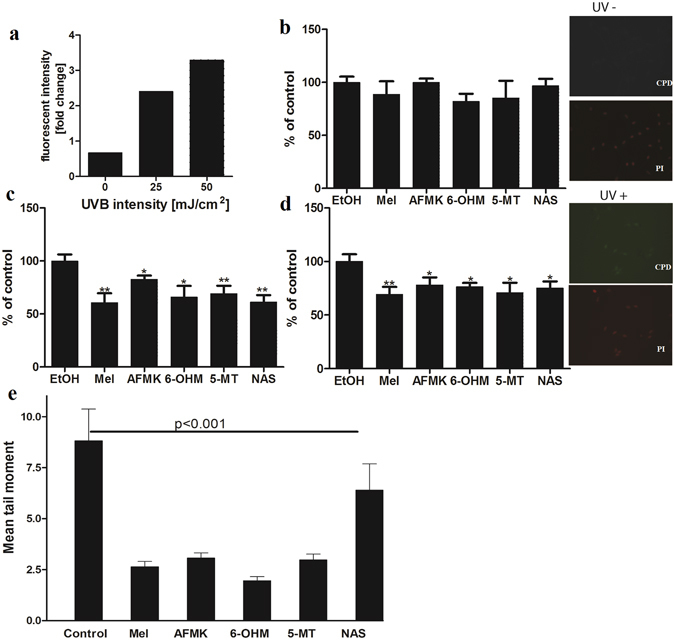
Melatonin, NAS, 6-OHM, AFMK, and 5-MT exhibit repair capacities in UVB-induced DNA damage in treated melanocytes as shown by reduced levels of CPD and comet cell tail moments. Cells were treated with melatonin or its metabolites for 24 h prior UV irradiation at the concentration 5 × 10⁻⁵ M, further exposed to UVB intensities of 25 or 50 mJ/cm2 and immediately treated again with melatonin or its derivatives for 3 h. Cells were fixed and stained with anti-CPD antibody and further imaged with a fluorescence microscope. Fluorescence intensity was analysed using ImageJ software and data were analysed using Graph Pad Prizm. CPD formation under different UVB intensity levels are shown in (a). Figure presents CPD levels after 0 mJ/cm2 (b), 25 mJ/cm2 (c) and 50 mJ/cm2 (d). Data are presented as percentile of control and were analyzed using t- test, *p < 0.05, **p < 0.01. As an example, images of the melanocytes stained with CPD antibody (green) and nuclear staining with propidium iodine (red) show no CPD signal in non-irradiated melanocytes (UV−) and positive CPD signal in UVB (25 mJ/cm2) irradiated cells (UV+) (magnification 20x). Also, melanocytes were treated with melatonin or its metabolites before and after UV irradiation (200 mJ/cm3). Tail moment is an index of DNA damage. Data are analysed using student t-test and p < 0.001 for all conditions (e).