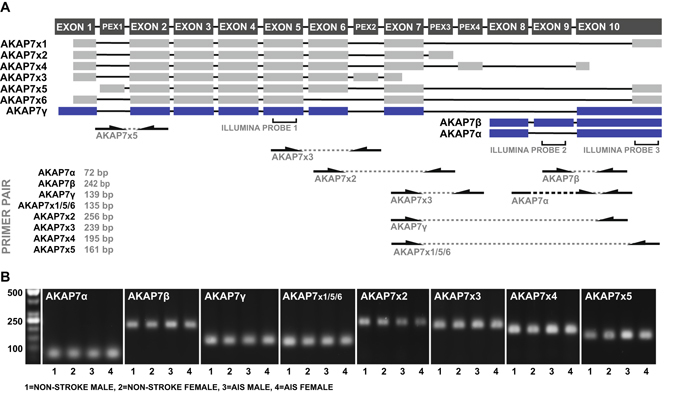
Figure 4.

Detection of AKAP7 splice variants in peripheral blood. (A) Exon maps of previously validated (blue) and bioinformatically predicted (grey) AKAP7 splice variants with the location of microarray probes and variant-specific priming sites. PEX denotes a bioinformatically predicted exon. (B) AKAP7 splice variant-specific RT-PCR products amplified from peripheral blood-derived cDNA obtained from two non-stroke donors and two stroke patients.
