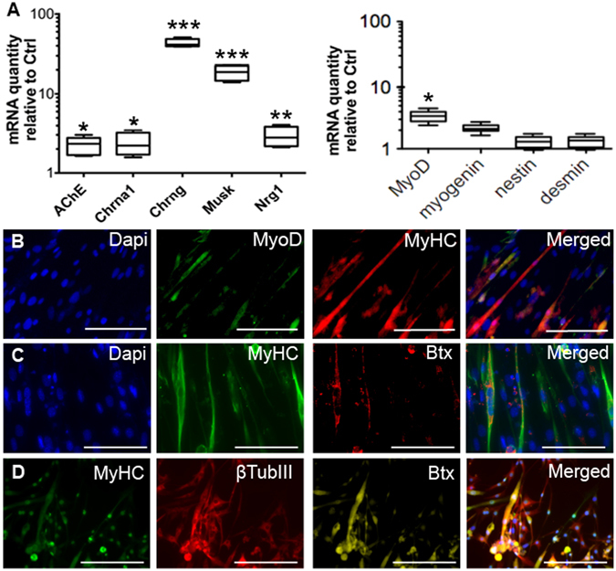
Figure 3.

Myogenic differentiation of iMuSCs and NMJ formation. (A) Whisker plots summarize the expression of NMJ-related genes as well as the myogenic genes within the neurosepheres in culture, analyzed by qPCR. (B) After the neurospheres were replaced into monolayer culture in MD Medium, their recovered their myogenic ability, formed myotube-like structures, and begun expression of myogenic protein MyoD (green, B), MyHC (Red, B; green, C,D). The attached neurospheres also formed neuron-like structures expressing β-Tubulin III (red, D). The presence of NMJs was shown by Btx staining (red, C; yellow, D). Nuclei were stained with Dapi. Scale bar = 100 µm.
