Figure 5.

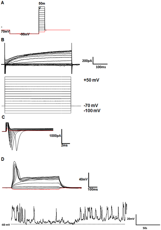
Electrophysiological evidence for successful neuronal development. (A) Whole-cell voltage clamp recording protocol used for patch-clamp analysis. (B) Representative example for K+ currents observed during 500 ms voltage steps of the whole-cell voltage clamp recording protocol displayed in (A). (C) Representative example of Na+ currents observed during 20-ms voltage steps of the whole-cell voltage clamp recording protocol displayed in (A). Note that Na+ currents (downward deflections) were observed at voltages ≤−30 mV. (D) Individual traces of action potential-like events generated by an intracellular current injection protocol. Spontaneous action potential-like events were recorded in current-clamp mode (0 pA). The dashed line indicates the −60 mV resting membrane potential.
