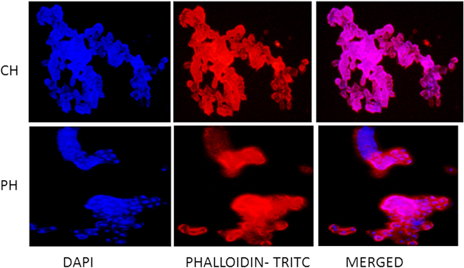
Figure 4.

Fluorescent imaging of the primary buffalo hepatocytes cultured on collagen-coated and polyHEMA-coated plates on the sixth day at 200x magnification. The nucleus of the primary buffalo hepatocytes were stained with DAPI and the cytoskeletal protein F-actin was stained with Phalloidin-TRITC stain. Hepatocytes formed patches on collagen-coated plates (CH) on the sixth day of the culture. However, different layers of cells could be observed in the intact spheroids formed on the polyHEMA coated dishes on the sixth day (PH).
