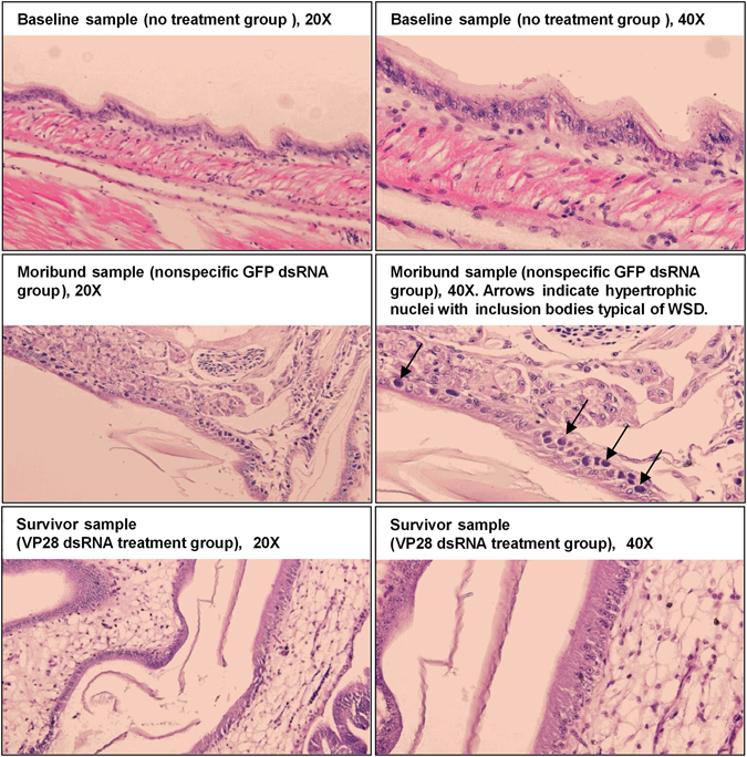
Figure 3.

Histology examination carried out to identify WSSV-specific pathology in experimental animals. Shrimp collected at different time points were sectioned and stained with hematoxylin-eosin for histological examination. All baseline samples were collected from the no treatment (PBS) group prior to challenge at 0 dpi, and all survivor samples were collected at 10 dpi from the VP28 dsRNA treatment group. Moribund shrimp were collected from the GFP dsRNA treatment group during the challenge trial, where infected cells exhibiting hypertrophic nuclei containing eosinophilic inclusion bodies were identified (indicated by black arrows).
