Figure 3.

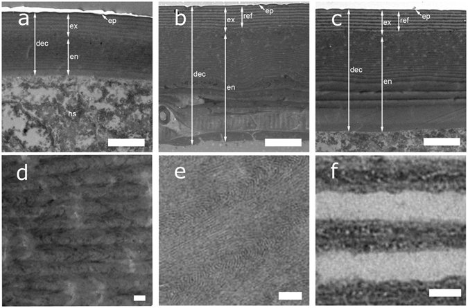
Figure 3 TEM sections of the developing dorsal elytral cuticle (“dec” in the figure), which illustrate the development of the endocuticle (“en”), exocuticle (“ex”), and epicuticle (“ep”). The cuticle after ecdysis (Stage 4: (a)) is considerably thinner than in the later stages 5 and 6 (b,c). The hemolymph space (“hs”) is visibile underneath the elytra. In (b), the young imago shows a more defined exocuticular multilayer reflector (“ref”) and a thicker endocuticle. The old imago, (c) exhibits a fully-formed cuticle. At higher magnification, it is possible to appreciate the structure of the inner exocuticle in (d,e) and the alternating fibril arrangement in the multilayer reflector in (f) for the fully formed imago. Scale bar: 2 μm for (a–c), and 100 nm for (d–f).
