Figure 9.

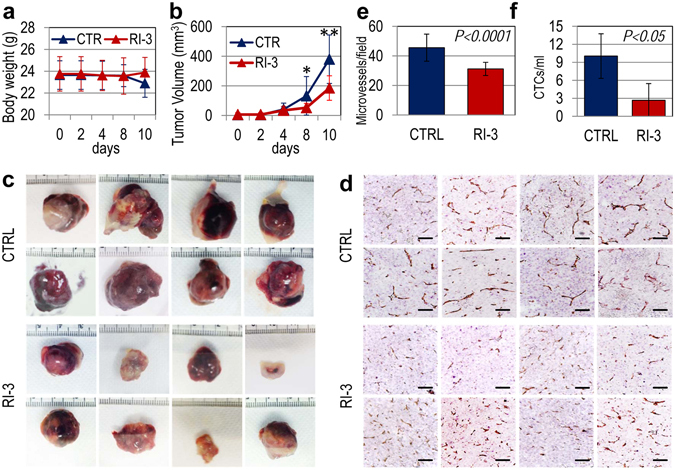
RI-3 reduces tumor growth, intra-tumoral microvessel density and vascular infiltration by sarcoma cells. Sixteen six-eight weeks old, Foxn1nu/nu female nude mice of 23 to 25 g, received an injection of Sarc cells into the right flank as a single-cell suspension (1 × 10⁶ cells in 100 µl of sterile PBS, 96% viability). Eight animals received i.p. injection of 6 mg/kg peptide RI-3 every 24 hr and eight received injections of vehicle only (CTRL). After 10 days, mice were anesthetized and blood samples (~500 µL/mouse) from the retroorbital venous plexus of mice were collected prior to sacrifice by CO2 inhalation. (a) Body weight measured every 2 days. (b) Tumor volumes measured by caliper every 2 days using the formula ½ × (width)2 × length (mm), where the width and the length are the shortest and the longest diameters of each tumor, respectively. (c) Excised tumors from vehicle-(CTRL) and RI-3-treated mice. (d) Tumors were fixed in buffered formalin, processed for paraffin sectioning and the sections immuno-stained with CD31. (e) Microvessel density was assessed by counting CD31 positive vascular channels in 5 randomly chosen fields per section, in at least two sections per tumor at × 200 magnification. (f) DNA from nucleated cells of murine blood samples was extracted and quantitated by Real-Time PCR using primers targeting Alu-sequences. Number of CTCs was calculated by comparing the obtained amplification curves with others generated in spiking experiments which were included in every run.
