Figure 2.

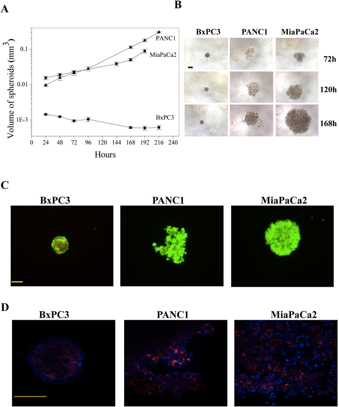
In vitro cell growth characteristics of PDAC cells in 3D culture conditions. (A) Comparison of growth rate between the PDAC cell lines with respect to difference in spheroid volume per day (n = 3). Data are means ± SEM of three separate experiments each carried out in triplicate. At the 0.05 level, data relative to MiaPaCa2 and PANC1 are not significantly different, whereas data relative to MiaPaCa2 are significantly different from those relative to BxPC3 (p = 0.003), and data relative to PANC1 are significantly different from those relative to BxPC3 (p = 0.002). (B) Microscopic images of PDAC at 72 h (top layer), 120 h (middle layer) and 168 h (bottom layer). (C) Live and dead cells were stained after 72H of culturing by Calcein (green) and PI (red) respectively. (D) Cytoskeleton organisation was studied by actin staining in all the three cell lines found to be on the periphery of cells. Scale bars represent 100 µm.
