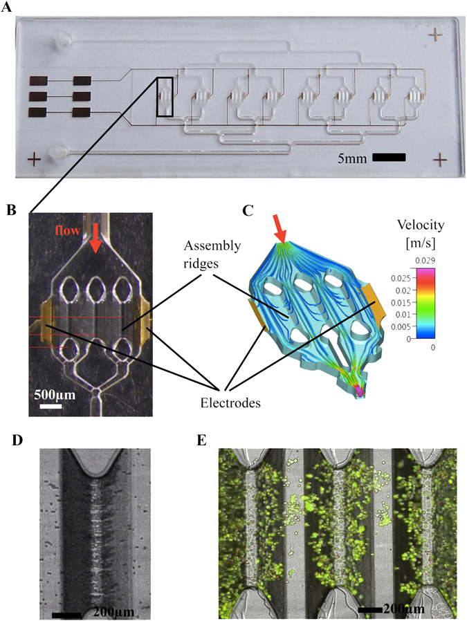
Figure 3.

HepaChip: (A) Full view of the chip with 8 culture chambers, fluidic inlet and outlet and gold electrodes. (B) close up of one chamber containing 2 electrodes and 3 assembly ridges coated with collagen. (C) Simulation of flow velocity and trajectories of cells during DEP assembly inside a culture chamber. (D) PANC1 cells assembled on one assembly ridge right after assembly. (E) Live/Dead staining of PANC1 cells after 146 hours of perfused culture inside the HepaChip® chamber.
