
Figure 1.
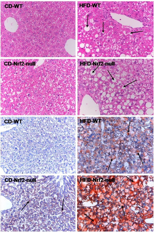
Mouse hepatic pathology. Liver sections of the former four figures were stained by hematoxylin and eosin. Liver sections of the latter four figures were stained with Oil Red O and the fat accumulated in hepatocytes manifested red. All the liver sections were examined by light microscopy and the images were displayed at 200× the original magnification. The fat accumulation in hepatocytes were pointed out by the arrows in HFD groups.