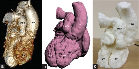
Figure 1.

The conversion of imaging data from patient 1 into its three-dimensional print. (a) Volume rendered gadolinium-enhanced three-dimensional magnetic resonance angiogram depicting spatial anatomy of the ventricles and the great arteries. (b) The hollow heart virtual three-dimensional model generated after postprocessing of the original imaging data. (c) The three-dimensional model printed in polyamide material using SLS technique. These images show that the three-dimensional model is identical to the original anatomy. Ao: Aorta, LV: Left ventricle, MPA: Main pulmonary artery, RV: Right ventricle, SLS: Slow laser sintering
