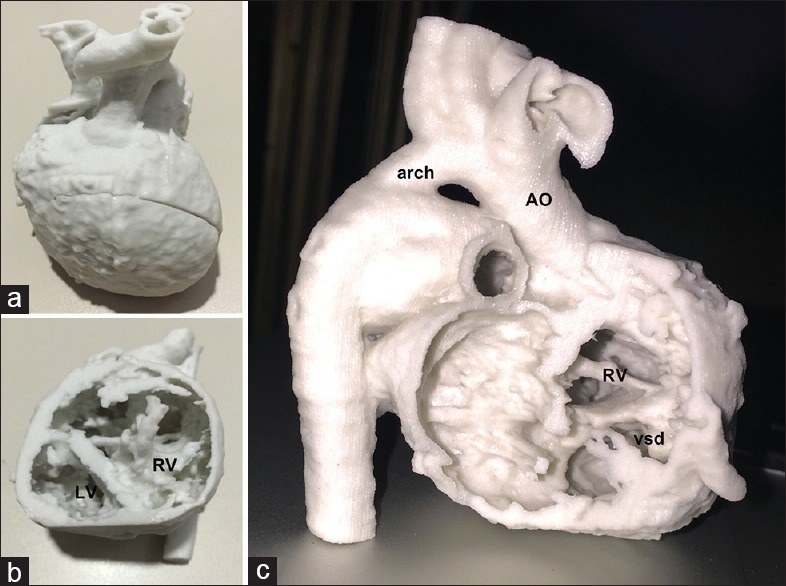
Figure 4.

(a-c) The three-dimensional print of patient 3 (criss-cross heart, double-outlet right ventricle), printed using “sandstone” material. (a) The “whole heart” perspective. (b) The view into the heart after removing the apical part of the model. (c) The transatrial perspective. Ao: Aorta, arch: Aortic arch, RV: Right ventricle, vsd: Ventricular septal defect
