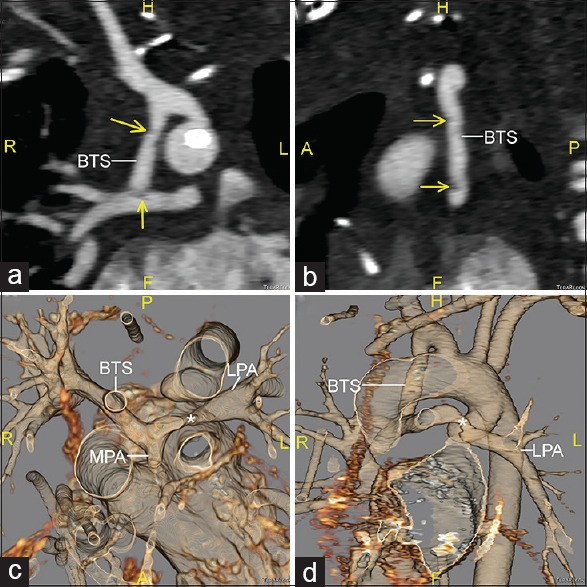
Figure 1.

Computed tomographic angiography images reconstructed in oblique coronal (a) and sagittal (b) planes, along with 3D volume-rendered reconstructions viewed from superior (c) and anterior-leftward (d) perspectives. The Blalock–Taussig shunt is seen inserting into the right pulmonary artery. Small thrombi are seen in the mid-portion of the shunt and its distal insertion into the right pulmonary artery (arrows). There is a small main pulmonary artery stump present, and an area of focal stenosis is seen in the left pulmonary artery (*). BTS: Blalock–Taussig shunt; RPA: Right pulmonary artery; LPA: Left pulmonary artery; MPA: Main pulmonary artery
