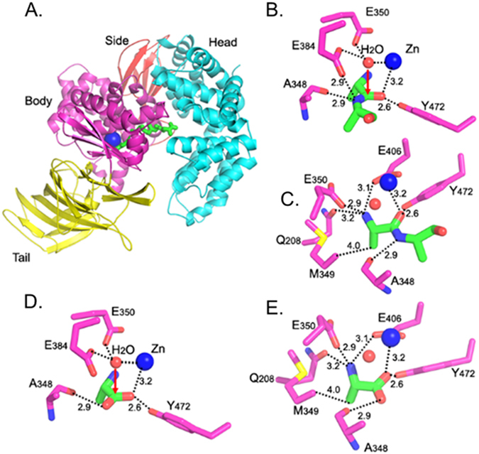
Figure 2.

Catalytic mechanism of pAPN. (A) Overall structure of pAPN complexed with a peptide substrate (PDB 4FKF). pAPN contains four domains: head (in cyan), side (in brown), body (in magenta), and tail (in yellow). Zinc is shown as a blue ball, and the peptide substrate is in green. (B) Interactions between catalytic residues of pAPN (in magenta) and the scissile peptide bond of the peptide substrate (in green). Catalytic water is shown as a red ball. (C) Another view of the structure in panel (B) to show all of the interactions between pAPN and the N-terminal residue of the peptide substrate. (D) Interactions between catalytic residues of pAPN (in magenta) and product of APN catalysis - free alanine (in green) (PDB 4FKH). (E) Another view of the structure in panel (D) to show all the interactions between pAPN and free alanine.
