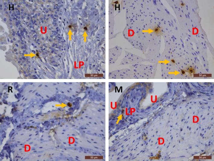
Figure 3.

Immunohistochemical stains for MCT on the bladder in human beings (upper panels) and mouse and rat (lower panels). In human bladder, MCT + mast cells are frequently observed, while in mouse and rat bladder, mast cells are sparse (yellow arrows). H, human; R, rat; M, mouse; U, urothelium; LP, lamina propria; D, detrusor. Scale bars: 50 μm.
