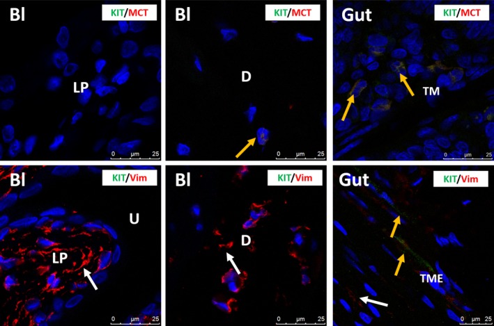
Figure 4.

Upper panels: confocal immunofluorescence double staining for KIT (green) and MCT (red) showing co‐occurrence of both antigens on a mast cell in the detrusor of rat bladder (yellow arrow). Mast cells are very rare in rat bladder. Gut tissue is used as external tissue control, showing KIT +/MCT + mast cells (yellow arrows) in the tunica mucosa (TM). Lower panels: confocal immunofluorescence double staining for KIT (green) and VIM (red) showing many VIM +/KIT − IC (white arrows) in rat bladder. Gut tissue is used as external tissue control, with the presence of KIT +/VIM + ICC (yellow arrows) and KIT −/VIM + IC (white arrow) in the tunica muscularis externa (TME). Bl, bladder; LP,lamina propria; D, detrusor; TM,tunica mucosa; TME, tunica muscularis externa. DAPI blue stain illustrates nuclei. Scale bars: 25 μm.
