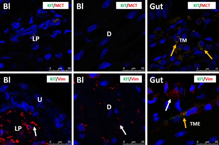
Figure 5.

Upper panels: confocal immunofluorescence double staining for KIT (green) and MCT (red), showing no immunoreactive cells in mouse bladder. Mast cells are very rare in mouse bladder. Gut tissue is used as external tissue control, showing KIT +/MCT + mast cells (yellow arrows) in the tunica mucosa (TM). Lower panels: confocal immunofluorescence double staining for KIT (green) and VIM (red) showing many VIM +/KIT − IC (white arrows) in mouse bladder. Gut tissue is used as external tissue control, with the presence of KIT +/VIM + ICC (yellow arrow) and KIT −/VIM + IC (white arrow) in the tunica muscularis externa (TME). Bl, bladder; LP, lamina propria; D, detrusor; TM, tunica mucosa; TME, tunica muscularis externa. DAPI blue stain illustrates nuclei. Scale bars: 25 μm.
