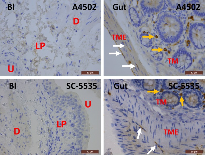
Figure 6.

Immunohistochemical stains for KIT on rat bladder and gut with clones A4502 (upper panels) and SC‐5535 (lower panels). In the bladder, almost no KIT + mast cells are observed, while in the gut, KIT is found on mast cells (yellow arrows) and ICC with long cytoplasmic processes (white arrows). Both antibody clones show similar expression patterns for KIT in the bladder and gut. Bl, bladder; U, urothelium; LP, lamina propria; D, detrusor; TME, tunica muscularis externa; TM, tunica mucosa. Scale bars: 50 μm.
