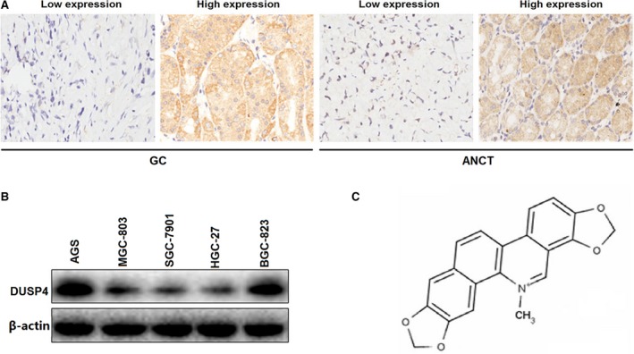
Figure 1.

The expression level of DUSP4 in GC tissues and cell lines. (A) Representative microphotographs of DUSP4 immunohistochemical staining in GC and ANCT tissues (×200). (B) The protein expression levels of DUSP4 in GC cell lines. (C) The chemical structure of sanguinarine.
