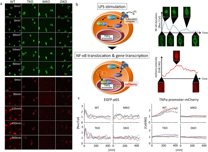
Figure 1.

The different roles of MyD88- and TRIF-dependent pathways in LPS-induced NF-κB nuclear translocation and TNFα promoter activation. (a) Live cell imaging of EGFP-tagged NF-κB nuclear translocation (upper panels) and TNFα promoter-driven mCherry fluorescence (lower panels) in wild-type (WT), TRIF−/− (TKO), MyD88−/−(MKO), and TRIF−/−/MyD88−/− (DKO) in response to 500 ng/mL LPS. The scale bar indicates 50 μm. (b) A schematic diagram of TLR4 signaling to NF-κB and TNFα promoter activation in the reporter cells. TLR4 signal is mediated by MyD88- and TRIF-dependent pathways to activate NF-κB. The activated NF-κB translocates into the nucleus where it associates with the TNFα promoter, which induces mCherry gene transcription. (c) Time course of the ratio of NF-κB in the nucleus (Nuc) to those in the cytoplasm (Cyt) (left column), and the ratio of mCherry fluorescence intensity inside a cell (Cell) to those outside (background (BG)) (right column) in the reporter cells. Each figure shows four representatives of approximately 50 cells from four independent experiments. Individual cells are shown by coloured lines.
