
Figure 4.
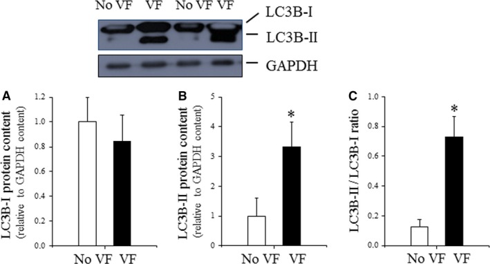
LC3B content in VF and non‐VF post‐IR heart tissue. (A) Cardiac LC3B‐I expression evaluated by Western immunoblotting and expressed relative to GAPDH content; (B) cardiac LC3B‐I expression evaluated by Western immunoblotting and expressed relative to GAPDH content. (C) Cardiac LC3B‐II/LC3B‐I ratio evaluated by Western immunoblotting (data are presented as n = 4 per group, *P < 0.05 No VF versus VF hearts.) Adapted from Meyer G, Czompa A, Reboul C, Csepanyi E, Czegledi A, Bak I, Balla G, Balla J, Tosaki A, Lekli I. The cellular autophagy markers Beclin‐1 and LC3B‐II are increased during reperfusion in fibrillated mouse hearts. Curr Pharm Des. 2013; 19 (39): 6912–8.