
Figure 1.
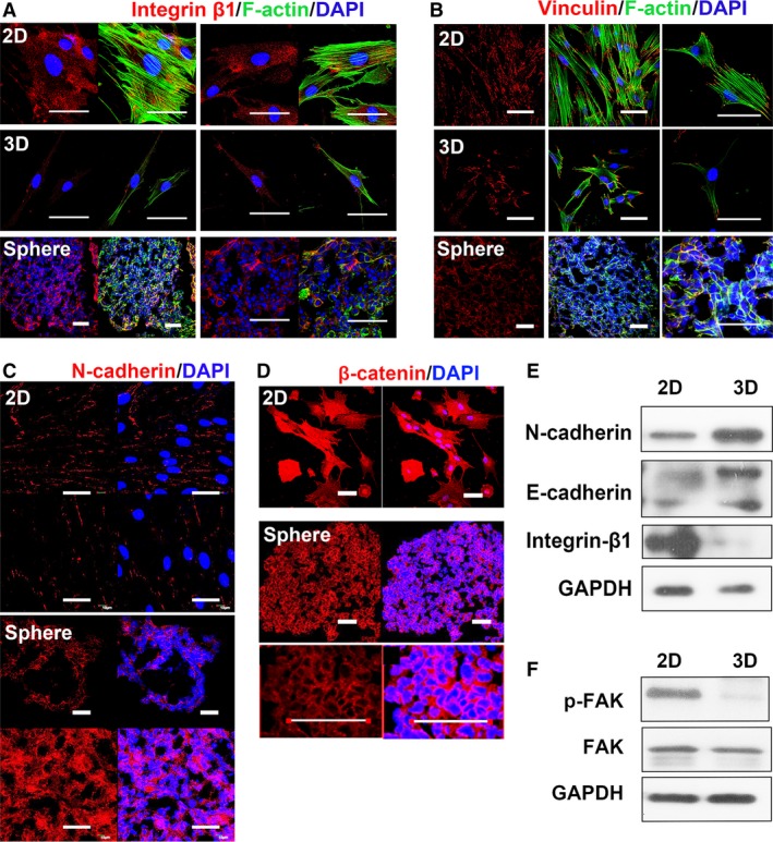
Adhesion molecules on the membrane of 2D and 3D MSCs. Membrane proteins in MSCs were detected with immunostaining for the indicated material. 2D: MSCs cultured as monolayer on slide, cultured overnight before fixation. 3D: MSCs, after 3D spheroid cultured for 60 hrs, trypsinized and re‐plated on slide, cultured overnight before fixation. 3D sphere: 10–20 μm of frozen section of fixed sphere, to analyse MSCs in situ. Immunostaining of integrin β1 (A) and Vinculin (B) were presented in red. F‐actin was labelled by phalloidin (Alexa‐Fluor 488‐conjugated, green). Cadherin‐based connection is established mostly at the cell–cell contact, so only confluent 2D MSC and 3D sphere were used to stain N‐cadherin (C) and β‐catenin (D). Red, antibody staining; blue, DAPI staining (scale bar, 50 μm). (E) Membrane fraction of 2D and 3D MSC were immunoblotted for N‐cadherin, E‐cadherin and integrin β1. Before membrane extraction, a fraction of cell from 2D and 3D MSCs were subjected to lysis. Immunoblotting for GAPDH with total cellular protein from this step was used as loading control. (F) Total cellular protein of 2D and 3D MSCs immunoblotting for FAK, phosphorylated FAK (p‐FAK) and GAPDH (loading control). These data are representative of five independent experiments with similar results.