
Figure 2.
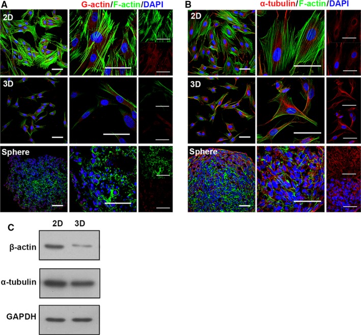
Cytoskeleton altered in 3D‐cultured MSCs. Two kinds of typical cytoskeleton in MSC were detected with immunostaining for the indicated material (abbreviation as in Fig. 1). (A) F‐actin was visualized using phalloidin (Alexa‐Fluor 488‐conjugated, green), G‐actin using DNase I (Rhodamine‐conjugated, red), higher magnification images were shown in right panel. (B) To show the structure of microtubule, α‐tubulin (red) was immunostained, higher magnification images were shown in right panel. Nuclei were labelled using DAPI (blue) (scale bar, 50 μm). (C) Total cellular protein of 2D, 3D MSCs immunoblotting for α‐tubulin, β‐actin, GAPDH (loading control). These data are representative of five independent experiments with similar results.