
Figure 3.
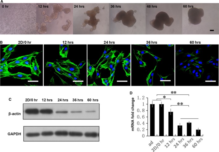
Actin‐cytoskeleton released gradually in 3D MSCs. (A) Microscope images show the process of sphere formation in 60‐hr duration. (B) Cultured in 3D for different duration, MSCs were trypsinized and re‐plated as monolayer and assessed for F‐actin arrangement with phalloidin (Alexa‐Fluor 488‐conjugated, green). Actin cytoskeleton restores the mechanism response in 3D culture, so MSCs derived from different time‐points in 3D culture presents continuous variation in F‐actin stress and arrangement (scale bar, 50 μm). Protein amount of β‐actin in 2D and 3D MSCs were analysed by Western blotting (C). These data are representative of three independent experiments with similar results. (A–C) Expression of β‐actin in 2D and 3D MSCs were analysed by real‐time PCR (D). Data are mean ± S.E.M. (n = 3); **P < 0.01 (Duncan's multiple range test).