
Figure 4.
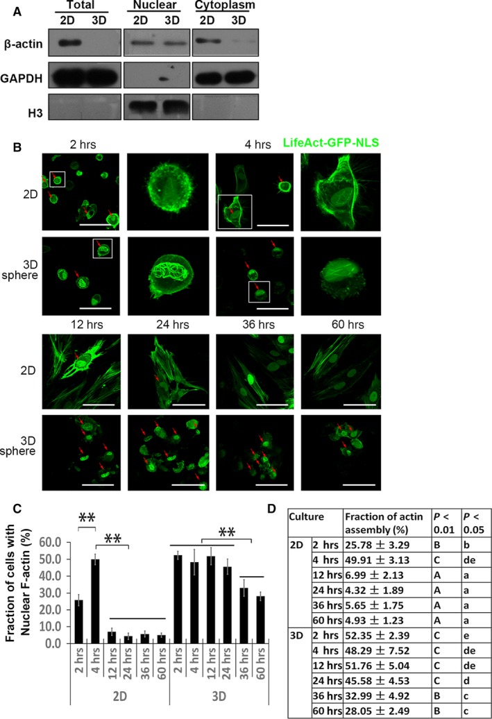
Temporal characterization of nuclear F‐actin formation during 2D and 3D culture. (A) Subcellular fractions were immunoblotted for β‐actin, GAPDH (cytoplasm loading control) and histone H3 (nuclear loading control). LifeAct‐GFP‐NLS was transfected to visualize nuclear F‐actin formation. MSCs were plated on culture dish as monolayer or dispersed in suspension to form spheres spontaneously and were monitored over time. Individual frames show F‐actin assembly at indicated time‐points (B). Red arrowheads mark MSCs with nuclear F‐actin formation. Higher magnification images of selected MSCs with typical nuclear F‐actin were shown. Quantification of nuclear F‐actin formation during culture duration as a fraction of cells with nuclear F‐actin over GFP positive cells (C and D) (scale bar, 50 μm). Four experiments were independently performed, and each time, over 50 cells were counted for each time‐point. Uppercase letters refer to P < 0.01, lowercases refer to P < 0.05 (Duncan's multiple range test).