
Figure 7.
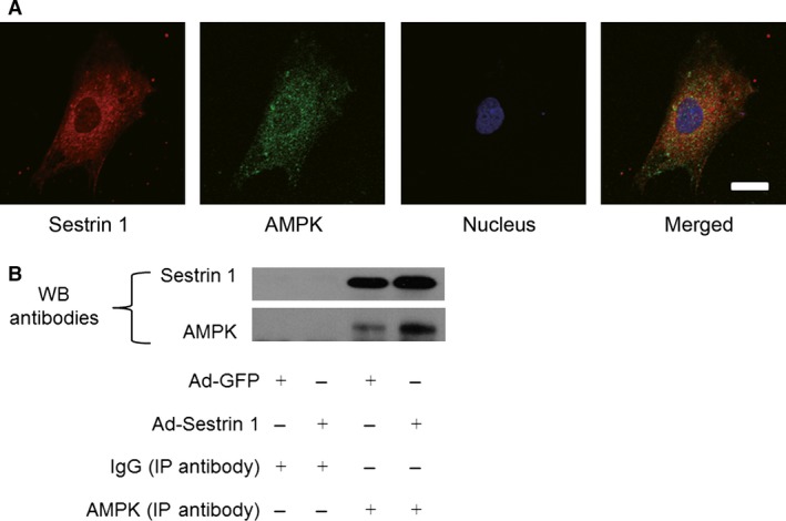
Co‐localization and interaction between Sestrin 1 and AMPK. (A) Immunofluorescence microscopy showing co‐localization of Sestrin 1 (red) and AMPK (green) in cardiomyocytes. Scale bar: 50 μm. (B) Co‐immunoprecipitation using AMPK antibody or rabbit normal IgG (negative control) in the lysates of cardiomyocytes transfected with Ad‐GFP or Ad‐Sestrin 1. Representative immunoblots with anti‐Sestrin 1 and anti‐AMPK were shown. Each of the experiments was repeated three times, n = 3.