Figure 2.

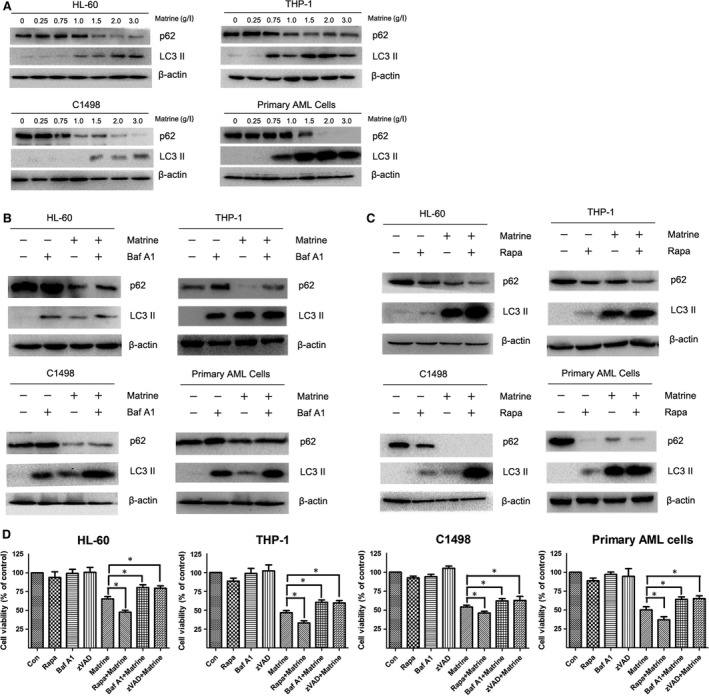
Matrine induces autophagy in AML cells. (A) HL‐60, THP‐1, C1498 and primary AML cells were treated with 0, 0.25, 0.75, 1, 1.5, 2, 3 g/l matrine for 24 hrs, and the autophagy markers LC3 II and SQSTM1/p62 were detected by Western blot analysis. (B ‐ C) AML cells were treated with 1.5 g/l matrine in the presence or absence of 10 nM bafilomycin A1 (Baf A1) or 20 nM rapamycin (Rapa) for 24 hrs, and the levels of LC3 II and SQSTM1/p62 were assessed by Western blot analysis. (D) After treatment with 1.5 g/l matrine in the presence or absence of 10 nM Baf A1 or 20 nM rapamycin or 10 μM z‐VAD‐FMK (zVAD) for 24 hrs, the cell viability was measured by CCK‐8 assay. Results were expressed as mean ± S.E.M. representing at least three independent experiments. *P < 0.05, versus matrine alone group.
