Figure 7.

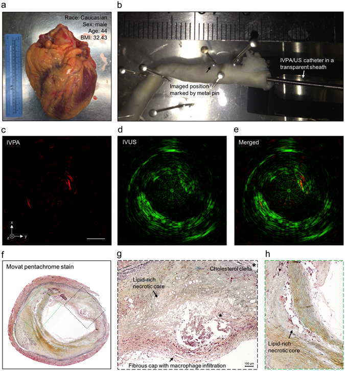
IVPA-US imaging of human coronary atherosclerosis at 16 fps with comparison to histopathology. (a) Picture of collected human heart. (b) Scenario picture of ex vivo IVPA-US imaging of dissected human coronary artery. The region of interest was marked by metal pin. The catheter and sheath were inserted into the artery lumen. Cross-sectional (c) IVPA, (d) IVUS, and (e) merged images of human coronary artery at the region of interest. (f) Gold-standard histopathology stained with Movat’s pentachrome at the region of interest. (g,h) Magnified images of lipid deposition sites corresponding to the dashed boxes in (f). *Indicates the accumulation of cholesterol clefts.
