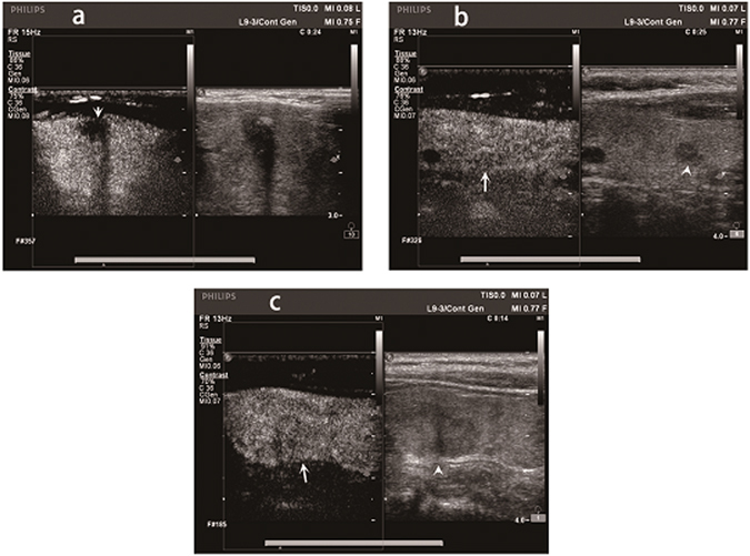
Figure 1.

The Contrast-enhanced ultrasound (CEUS) images of different papillary thyroid carcinoma (PTC) patients with extracapsular extension (ECE). (a) CEUS image of a 48-year-old male PTC patient with ECE. The CEUS image clearly showed that the anterior thyroid capsular was invaded by PTC (arrow). (b) CEUS image of a 58-year-old female PTC patient with ECE. The nodule showed by arrow head was the PTC nodule. ECE was not clearly confirmed by CEUS (arrow). (c) CEUS image of a 50-year-old female PTC patient with ECE. The nodule showed by arrow head was the PTC nodule. ECE was not clearly detected by CEUS (arrow).
