
Figure 2.
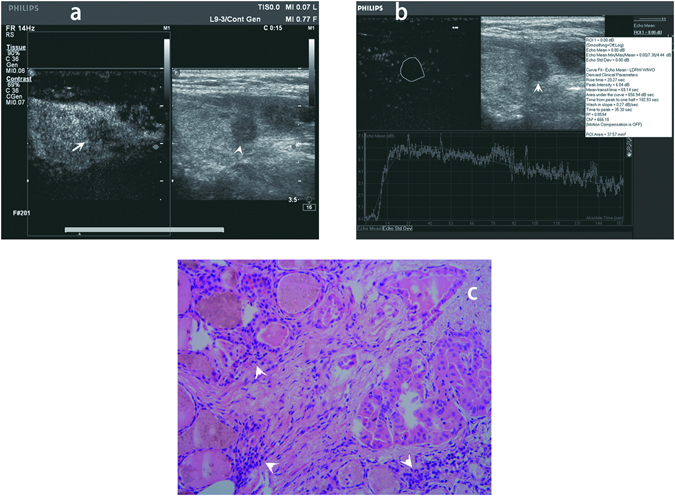
Qualitative and quantitative contrast-enhanced ultrasound (CEUS) image and histopathologic image of a 57-year-old female papillary thyroid carcinoma (PTC) patient without extracapsular extension (ECE). (a) Qualitative CEUS image of the patient. The PTC nodule in gray scale ultrasound image was showed by arrow head. The PTC nodule in CEUS image was showed by arrow. (b) Quantitative CEUS image of the patient. Time-intensity curve showed rise time 20.27 sec, peak intensity 6.04 dB, area under the curve 656.94dBsec, time from peak to one half 102.93 sec, wash in slope 0.27dBsec, time to peak 35.30 sec. (c) Histopathologic image revealed PTC of the patient without ECE. The cancer cells were showed by arrow head (original magnification, x100).