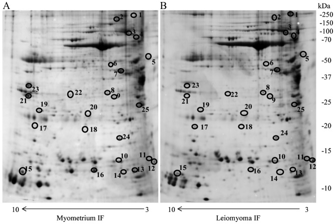
Figure 1.
Two-dimensional electrophoresis map of the (A) normal myometrium IF and (B) leiomyoma IF proteome. Immobilized pH gradient pH 3–10 non-linear strips were used for the first dimension and 12% polyacrylamide gels were used for the second dimension. IF, interstitial fluid. Circles indicate different proteins identified.

