
Figure 3.
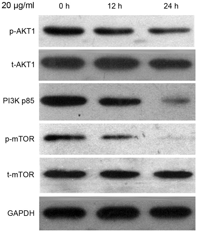
The protein expression level of PI3K p85, p-AKT1, t-AKT1, p-mTOR and t-mTOR were detected using western blot analysis. Ishikawa cells were treated with 20 µg/ml CDDP for 12 and 24 h, followed by harvesting for western blot analysis. PI3K, phosphoinositide 3-kinase; p-, phosphorylated; t-, total; AKT, protein kinase B; mTOR, mammalian target of rapamycin; CDDP, cisplatin.