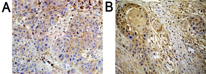
Figure 1.
Results of immunohistochemistry. Positively expressed SATB1 located in the nucleus with pale yellow-brown or brown color. In addition, (A) compared with well-differentiated tissues, (B) the expression of SATB1 was significantly higher in poorly differentiated tissues. (A) Well-differentiated; (B) poorly differentiated. Magnification, 200x.

