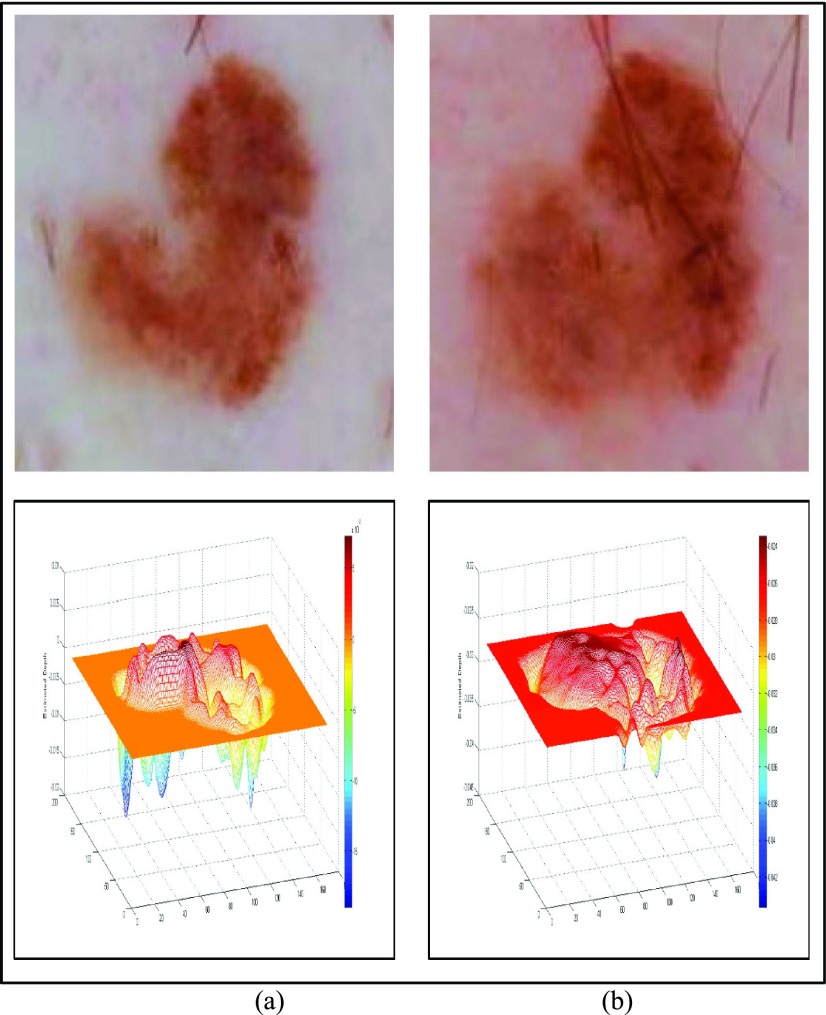
FIGURE 8.
Slow growing melanoma follow-up image from [61] and 3D depth projections (a) Baseline image (top) and estimated depth (bottom). (b) Follow-up image obtained after 5 years. Biopsy reveals melanoma is 0.15mm thick (top) and estimated depth (bottom).

