
Fig. 4. EMC divisions are symmetric for Prospero distribution but asymmetric for cell polarity and Notch signaling.
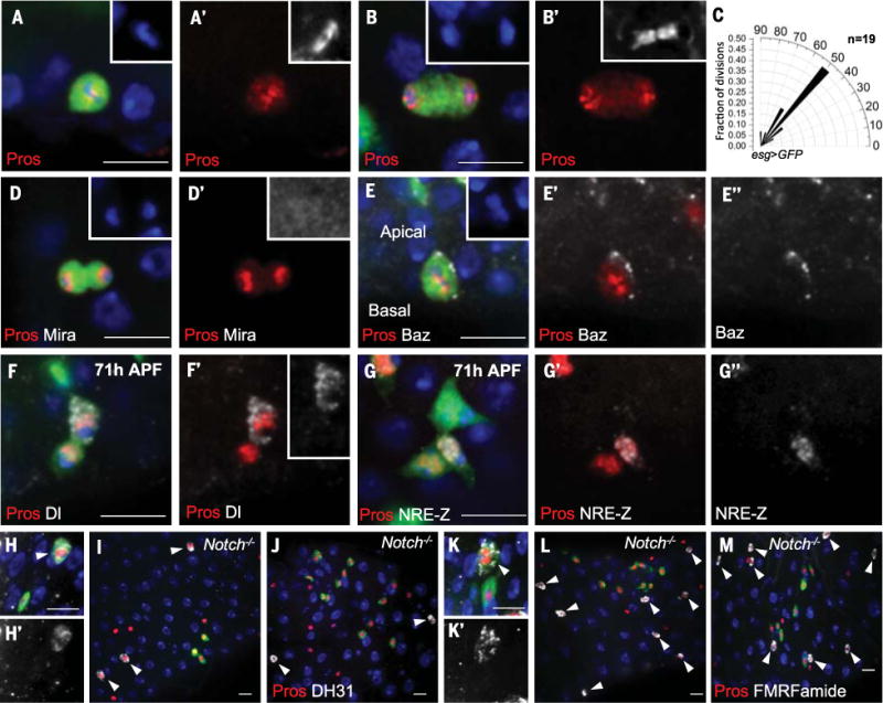
(A and B) Pros symmetrically localizes to both daughters during EMC metaphase (A) and telophase (B). Insets in (A) and (B), DAPI; insets in (A′) and (B′), α-tubulin. (C) Radial histogram quantification of division angles in metaphase of esg>GFP EMC mitosis. (D) Mira staining is absent during EMC division. Inset in (D), DAPI; inset in (D′), Mira (enhanced levels). (E) Baz staining (E″) localizes as a crescent on the apical cell membrane during EMC metaphase. Inset in (E), DAPI. (F) After EMC division (71 hours APF), strong Dl staining [inset in (F′)] is present in one of the Pros+ pair of cells. (G) After EMC division (71 hours APF), one of the two Pros+ cells is NRE-lacZ+ (G´´). (H to M) Wild-type [(H), (H´), (K), and (K´)] and Notch mutant [(I), (J), (L), and (M)] MARCM clones were induced at 24 hours APF, and midguts were dissected at 92 hours APF. In (H) and (H′), the peptide hormone DH31 (arrowhead) is present in a wild-type MARCM ee clone cell (green). Red, Pros; white, DH31. In (I) and (J), Notch mutant MARCM clones (green) are Pros+ but fail to stain for DH31. Arrowheads denote all DH31+ cells (white). [(K) and (K′)] The neuropeptide motif FMRFamide is present in a wild-type MARCM ee clone cell (green). Red, Pros; white, FMRFamide. [(L) and (M)] Notch mutant MARCM clones (green) are Pros+ but fail to stain for FMRFamide. Arrowheads denote all FMRFamide+ cells (white). Scale bars, 10 μm.