
Figure 2.
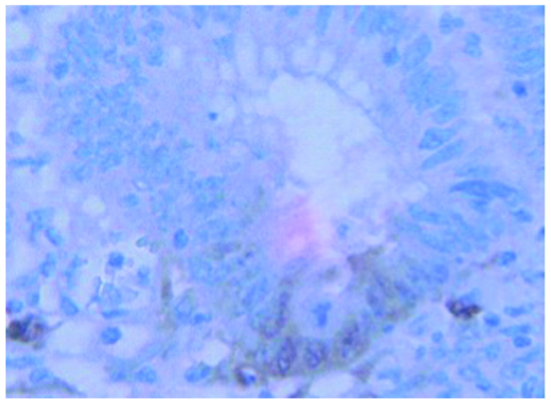
Cells that revealed immunohistochemical expression of IDO were considered positive. The positive cancerous cells were recognized in the submucosal layer. The case illustrated in this figure is T1 case. IDO, indoleamine 2,3-dioxygenase.
Official websites use .gov
A
.gov website belongs to an official
government organization in the United States.
Secure .gov websites use HTTPS
A lock (
) or https:// means you've safely
connected to the .gov website. Share sensitive
information only on official, secure websites.
Cells that revealed immunohistochemical expression of IDO were considered positive. The positive cancerous cells were recognized in the submucosal layer. The case illustrated in this figure is T1 case. IDO, indoleamine 2,3-dioxygenase.