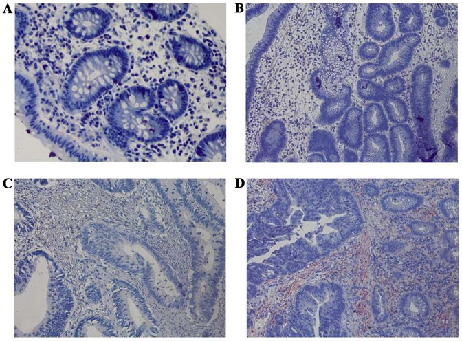
Figure 4.
Immunohistochemical staining of FasL among the four groups at a magnification of ×400. (A) Negative expression of FasL in normal colorectal mucosa. (B) Negative expression of FasL in colorectal adenoma. (C) Negative expression of FasL in early colorectal cancer. (D) Strong positive expression of FasL in advanced colorectal cancer. FasL, apoptosis antigen 1 ligand.

