
Figure 7.
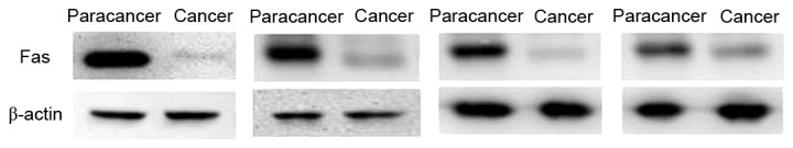
Expression of Fas in paracancer tissues and advanced colorectal cancer tissues. The expression of Fas was decreased in the advanced colorectal cancer group compared with the paracancer group. Staging and location of samples from left to right: Dukes C1, ascending colon; Dukes C1, rectum; Dukes C1, sigmoid; and Dukes B, rectum. β-actin was the internal reference. Fas, apoptosis antigen 1.