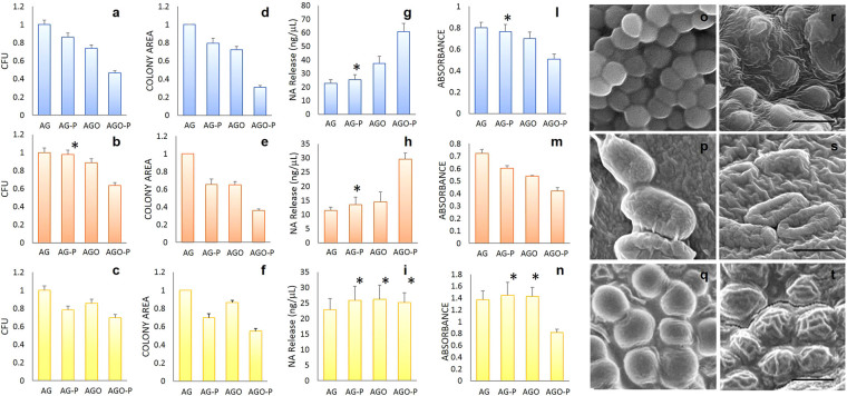
Figure 2.
CFUs number, colony size, cell damage, metabolic activity and structural integrity of microorganisms grown on different substrates. Number of CFUs on different hydrogels: S. Aureus (a), E. Coli (b) and C. Albicans (c). Normalized colony diameter on different hydrogels: S. Aureus (d), E. Coli (e) and C. Albicans (f). Nucleic acid released after exposure to different hydrogels of S. Aureus (g), E. Coli (h) and C.Albicans cells (i). Metabolic activity quantification using XTT test for S.Aureus (l), E. Coli (m) and C.Albicans cells (n). Representative Scanning Electron Microscopy images of for S.Aureus (o), E. Coli (p) and C.Albicans (q) on AGO hydrogels or AGO-P hydrogels (r–t). Scale bar is 1 μm in (r) and (s) and 10 μm in (t). Asterisks indicate statistically not significant differences compared to the untreated agar hydrogel AG.

