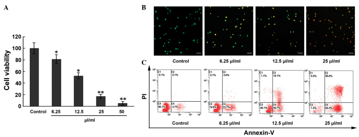
Figure 1.
Apoptosis and anti-proliferative effects induced by MTE on ECs. (A) ECs were treated with MTE (0, 6.25, 12.5, 25 and 50 µl/ml) for 48 h. The cell viability was analyzed by the 3-(4,5-dimethylthiazol-2-yl)-2,5-diphenyl-2H-tetrazolium bromide assay. (B) Following incubation with different concentrations of MTE (0, 6.25, 12.5, 25 µl/ml) for 48 h, the cells were stained with acridine orange/PI solutions and observed under a confocal laser scanning microscope. Green, jacinth and red spots represent living, apoptotic and necrotic cells, respectively (x200 magnification). Bar, 10 µm. (C) ECs were incubated with the indicated doses of MTE for 48 h. After being stained with Annexin V-fluorescein isothiocyanate and PI, apoptotic cell death (%) was determined with flow cytometry. Lower left, Annexin V−/PI− cells (normal); lower right, Annexin V+/PI− cells (early apoptosis); upper right, Annexin V+/PI+ cells (late apoptosis); and upper left, Annexin V−/PI+ cells (necrosis). Results are represented as the mean ± standard deviation from three separate experiments. *P<0.05;**P<0.001 vs. control. MTE. Marsdenia tenacissima extract; EC, endothelial cell; PI, propidium iodide.

