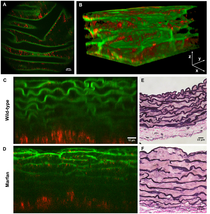
Figure 1.

Aortic tissue visualizations using multiphoton microscopy. (A) En-face perspective of the IEL of the ascending aorta from a WT mouse using multiphoton microscopy. TPEF signal of elastin in green and SHG signal of collagen in red. (B) Three-dimensional rendering of a portion of the tunica media from the ascending aorta of a WT mouse. (C and D) Representative multiphoton images of the tunica media in transverse views of WT (C) and Marfan (D) mice. (E and F) Conventional histological visualization of elastic fibres using Verhoeff-van Gieson staining in WT (E) and Marfan (F) aortic tissue. Scale bar, 10 μm.
