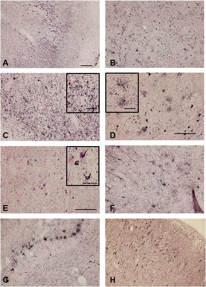
Figure 2.

Photomicrograph of phosphorylated tau immunohistochemistry of the temporal lobe of the GRN mutation cases. Massive AT8-positive structures were observed in the hippocampus (A), amygdala (B), inferior temporal cortex (C) in case 2. AT8-positive astrocytes were observed in the temporal cortex (D) and AT8-positive oligodendrocytes in the white matter of temporal lobe (E) in case 2. AT8-positive deposition was also detected in amygdala in case 1 (F), hippocampus in case 3 (G) and entorhinal cortex in case 4 (H). The sections were counterstained with Kernechtrot stain solution. The scale bar in A applies to (B,C,D,F,G and H) (200 μm), in E (100 μm), respectively. The scale bars in the inserts are 50 μm (C and D) and 25 μm (E), respectively.
