
Figure 3.
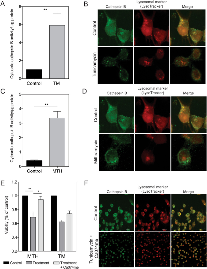
ER stress leads to a disruption in the lysosomal membrane. MIA PaCa-2 cells analyzed for lysosome membrane permeabilization. (A) Cells treated with 1 μM tunicamycin and analyzed for cytosolic cathepsin B activity. (B) Cells treated with 1 μM tunicamycin, and stained with cathepsin B and lysotracker. Images taken at 60x magnification. (C) Cells treated with 100 nM mithramycin and analyzed for cytosolic cathepsin B activity. (D) Cells treated with 100 nM mithramycin, and stained with cathepsin B and lysotracker. Images taken at 60x magnification. (E) Viability determined for cells treated with 100 nM mithramycin or 1 μM tunicamycin, with and without a cathepsin B inhibitor, Ca074me. (F) Cells treated with 1 μM tunicamycin, with and without Ca074me detected by immunofluorescence. Images taken at 40x and 60x magnification.