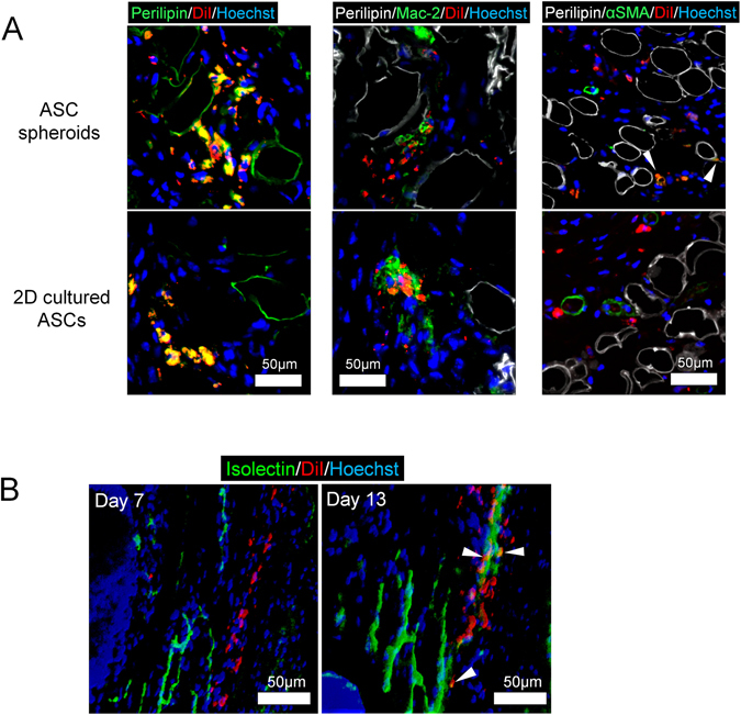
Figure 6.

Immunohistological tracing of administered ASCs in irradiated skin ulcers of nude mice. (A) Immunohistological views of irradiated ulcer specimens 13 days after surgery and transplantation for tracing of administered ASCs (DiI-labeled cells). Cell fates were visualized by immunostaining for perilipin, Mac-2, and αSMA. Scale bars = 50 µm. (B) Whole mount staining of the irradiated wound tissue at days 7 and 13. Vascular endothelial cells were labeled by isolectin. Scale bars = 50 µm.
