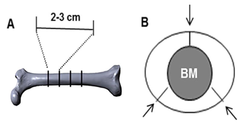
Figure 1. Illustration of transverse and segmental cuts of bovine femur.

A. An image of bovine femur depicting diaphyseal portions that are transversely cut into 2–3 cm cylinders; B. Bony cylinders referred in (A) are further cut into three segments as indicated (arrows).
