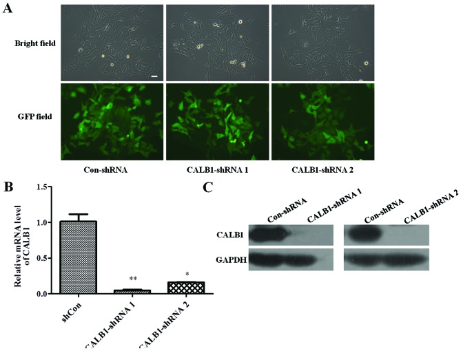
Figure 1.
Confirmation of CALB1-knockdown in osteosarcoma U2OS cells with CALB1-shRNA 1 and 2 infection. (A) Expression of GFP in infected U2OS cells was shown in bright-field images and by GFP. Scale bar, 10 µm. Images were captured at 72 h post-infection (magnification, ×100). (B) Quantitative polymerase chain reaction and (C) western blot analysis demonstrating that the CALB1 mRNA and protein levels were knocked down in CALB1-shRNA 1- and 2-infected cells. *P<0.05, **P<0.01. CALB1, calbindin 1; GFP, green fluorescent protein; sh, short hairpin; con, negative control.

