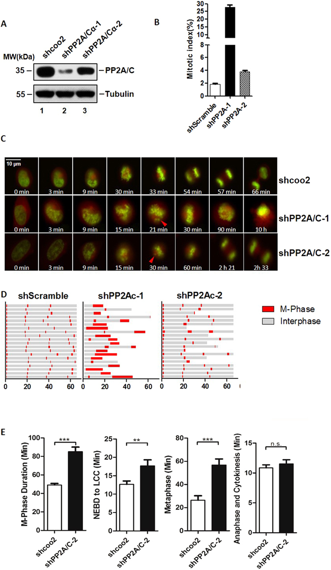
Figure 1.

PP2A knockdown leads to metaphase delay in HeLa cells. (A) Protein depletion levels of PP2A/C evaluated by Western blot in cells after 48 hours infection with indicated shRNAs. For the original uncropped western blot data, see Supplementary information. (B) Mitotic index (MI) of cells (A), was calculated by counting metaphase cells in approximately 1000 cells. (C) Snapshots of Hela cells (shcoo2, shPP2A/C-1, shPP2A/C-2) taken at indicated times (scale bar indicates 10 μm). Red triangles indicate misalignment which has occurred during metaphase. (D) Cell cycle progression of HeLa cells infected with lenti-viruses (shcoo2, shPP2A/Cα-1, and shPP2A/Cα-2). Cells were monitored by time-lapse fluorescence microscopy. H2B-GFP and DsRed-α-tubulin were stably expressed in HeLa cells to indicate chromatin status. Single cells were monitored for 65 hours. Gray sections on the progress bar represent interphase, and red sections represent mitotic phase. (E) Mean length of M-phase, NEBD-LCC (Nuclear envelope breakdown to last chromosome congress), Metaphase and anaphase in HeLa cells infected with shPP2A/C-2 and shcoo2 lenti-virus (N > 50).
