
Figure 1.
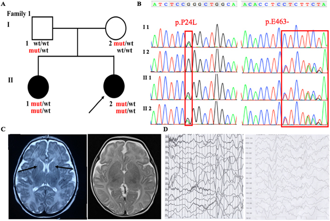
Clinical features of the patients and family 1 with the c.71 C > T, p.P24L; c.1387-1389delGAG, p.E463- in ADSL. (A) Pedigree structure of the studied family 1. In the family 1, WES was performed in I:1, I:2, II:2. Also, the compound heterozygous mutations were presented in the pedigree. (B) The PCR products were sequenced with the reverse primers (ADSL c.71 C > T, p.P24L; c.1387-1389delGAG, p.E463-). (C) The brain MRI shows the abnormal myelination of white matter in the proband (1-month old) (Left), and normal signal of the II 1 (Right) with T2-weighted. (D) EEG of the patients show the abnormal multifocal spike and ware wave in the proband (Left) and II 2 (Right).